
Hip Arthroscopy
Clinical Practice Guidelines
These guidelines were created in an effort to improve hip arthroscopy outcomes based on the best available evidence in the literature. They were developed and are maintained in a joint effort by the West Coast Hip Preservation Institute and our specialized research assistants. All guidelines are meant to be suggestions, as we always recommend individualized patient diagnosis and treatment in line with patient goals to provide the best possible care. The variety of topics cover common modern hip arthroscopy pathology and techniques.
Topics
-

Positioning
Patient positioning and table selection during hip arthroscopy is a crucial aspect that can improve surgical safety and efficiency.
-
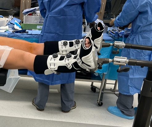
Traction
Traction is a necessary component of hip arthroscopy, but attention to appropriate tensioning is important to avoid complications.
-

Access
There are multiple ways to access the hip joint to perform surgery. There are advantages and disadvantages for each technique.
-

Labral Treatment
Treatment of labral pathology may involve debridement, repair, or reconstruction. Here we evaluate the literature comparing debridement and repair.
-

Segmental Labral Reconstruction
Segmental labral reconstruction is an option for labral pathology that only involves part of the labrum in cases where the labrum is beyond repair.
-

Circumferential Labral Reconstruction
When the entire labrum is irrepairable, circumferential labral reconstruction may be performed to replace the entire labrum.
-

Femoroplasty
Removal of a cam lesion, also known as femoroplasty, is a common procedure performed for patient with femoroacetabular impingement syndrome.
-

Capsular Repair
The hip capsule is routinely incised to perform surgery. These guidelines examine whether capsular repair is necessary.
-

Capsular Plication
Capsular plication, or tightening of the capsule, is a procedure that can be performed during arthroscopy for hip instability.
-
Open versus Arthroscopic Gluteal Repair
Evaluation of open versus arthroscopic approaches for gluteal tendon repair.
-
Open versus Arthroscopic Hamstring Repair
Evaluation of open versus arthoscopic approaches for hamstring repair
-
Iliopsoas Lengthening
Iliopsoas lengthening for contracture or symptomatic tendinosis after total hip arthroplasty